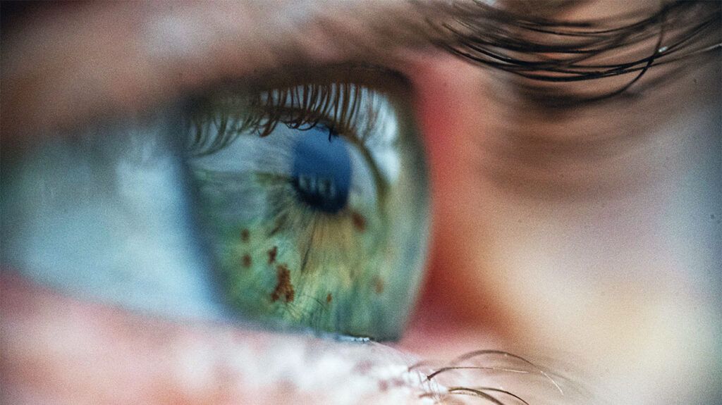
Retinal hemorrhage is the medical term for bleeding in the retina. There are various types of retinal hemorrhages, including vitreous, intraretinal, and subretinal bleeds.
The retina is a layer of light-sensitive cells in a person’s eye. These cells detect light and convert it into signals to pass to the brain, which uses these signals to create a visual picture.
There are different types of retinal hemorrhage, which vary depending on the location of the bleeding. A retinal hemorrhage is often a symptom of an underlying disorder, such as diabetes, high blood pressure, or trauma to the head or eyes. They can also occur due to direct trauma.

Below are the different types of retinal hemorrhage.
Vitreous hemorrhage
The vitreous is the gel-like fluid
Some health experts may also refer to a VH as a preretinal hemorrage. This type of bleed
A VH will often cause fresh blood clots to appear in the vitreous and may cause a person to experience floaters. Floaters are
Intraretinal hemorrhage
Intraretinal hemorrhages occur when blood
These hemorrhages are often dense and dark red, with a sharp outline. Medical professionals may refer to them as “dot and blot hemorrhages.”
Subretinal hemorrhage
The photoreceptor layer in the eye
Subretinal hemorrhages occur when blood accumulates beneath the RPE. They are often deep red, broader in shape than other types, and have diffuse margins.
Learn about other retinal problems.
Retinal hemorrhages are
Retinal hemorrhages that have the shape of a dot, blot, splinter, or flame do not threaten vision and are not a medical emergency. Treatment for retinal hemorrhages will usually involve treating their underlying cause.
However, if a person experiences problems with their vision or the bleeding is the result of trauma, it may be advisable to seek medical assistance.
Retinal bleeding
- Eye diseases: Eye diseases that can cause retinal bleeding include:
- age-related macular degeneration (AMD)
- polypoidal choroidal vasculopathy
- optic disc hemorrhages
- Diabetic retinopathy: This
refers to blood vessel damage within the retina that occurs due to diabetes. Diabetic retinopathy can lead to vision loss and blindness. - Hypertensive retinopathy: This occurs when high blood pressure
causes damage to the blood vessels in the eyes. It is a progressive condition that may lead to vision loss. - Retinal vein occlusions: Retinal vein occlusions occur when a blood clot
blocks veins that transport blood out of the retina. In rare instances, blood clots form in the arteries carrying blood to the retina, causing blurry vision. This usually only affects one eye. - Trauma: Head or eye trauma may cause retinal hemorrhages. For example, infants may develop neonatal retinal hemorrhages due to birth trauma.
- Anemia: Anemia is a medical condition that occurs when there are
not enough red blood cells in the body. A person may also develop anemia if the red blood cells they do have cannot carry oxygen throughout the body as well as usual. Symptoms include:- fatigue
- shortness of breath
- heart palpitations
- changes in skin color
- headaches
- Leukemia: Leukemia is the umbrella term for cancers of the
blood cells . Different types of leukemia affect different blood cells. Some leukemias are fast-growing, and some are slow-growing. - Acute bacterial endocarditis: This condition causes inflammation in the heart’s lining
due to a bacterial infection. Common symptoms include:- fever, chills, and night sweats
- anorexia, weight loss, and loss of appetite
- malaise
- headaches
- muscle aches and pains
- joint pain
- shortness of breath
- Sickle cell anemia: Sickle cell anemia is the term for a group of blood disorders
affecting hemoglobin, the protein that carries oxygen throughout the body. Sickle cell anemia can cause a person to experience:- jaundice (yellowish skin)
- extreme fatigue
- painful swelling of the hands and feet
- Ocular ischemic syndrome: This is a
visual function disorder caused by carotid artery stenosis. It can cause visual loss and eye pain. - Preeclampsia: Preeclampsia is sudden high blood pressure that occurs during pregnancy. It may cause symptoms such as:
- a persistent headache
- vision changes
- stomach pain
- nausea or vomiting
Symptoms of retinal bleeding vary depending on the type of retinal hemorrhage. However, some common symptoms of retinal bleeding may include blurry vision and the presence of floaters in vision.
Diagnostic signs an ophthalmologist can see
- disc-shaped
- flame-shaped
- boat- or “D”-shaped
- round with a white center
They may have a diffuse or sharp outline and can be deep or dark red.
Retinal hemorrhages often occur
During diagnosis, a doctor will want to determine the cause of the bleeding so they can treat this underlying condition.
They will usually begin by measuring a person’s:
They may then conduct further testing to determine the underlying cause.
A retinal hemorrhage that does not obscure or threaten a person’s vision
A doctor will observe the hemorrhage during a follow-up appointment and track its progression and size. They will aim to diagnose the underlying condition that caused the hemorrhage and devise a treatment plan to treat this condition.
In some cases, a doctor may recommend the following treatment strategy for more serious hemorrhages:
- bed rest with head elevation to reduce further bleeding
- wearing an eyepatch on the affected eye to reduce eye movement
- avoiding using certain drugs, such as aspirin
In some cases, a medical professional may use antivascular endothelial growth factor to help slow or stop bleeding. They may also use laser photocoagulation or cryotherapy to close breaks in the retina.
A person with a detached retina may undergo surgery to reattach it.
Effective
A person may also wish to follow
- Undergo a comprehensive dilated eye exam: Medical professionals can check for signs of eye conditions during this procedure. This may help detect issues early on when they are easier to treat.
- Lifestyle changes: Certain conditions can increase a person’s risk of retinal hemorrhages. These include diabetes and high blood pressure. The following can lower the risk of developing these conditions:
- eating a balanced diet
- being physically active
- quitting smoking, if applicable
- Protect the eyes: A person may wish to follow these steps to protect their eyes:
- wear sunglasses
- wear protective eyewear when doing certain activities, such as playing sports or doing construction work
- regularly rest the eyes if looking at a computer screen for long periods
- wash hands before putting in or taking out contact lenses
A retinal hemorrhage occurs when there is bleeding in the retina. Retinal hemorrhages are often a sign that an underlying condition is present. Types of retinal hemorrhages include vitreous, preretinal, intraretinal, and subretinal hemorrhages.
Certain medical conditions can cause retinal hemorrhages. These include diabetes, high blood pressure, retinal vein occlusions, trauma, anemia, leukemia, and acute bacterial endocarditis.
In many cases, retinal hemorrhages do not require treatment. Instead, medical professionals often diagnose and treat the underlying condition that caused the hemorrhage. In some cases, a doctor may suggest laser photocoagulation or cryotherapy to close breaks in the retina.
