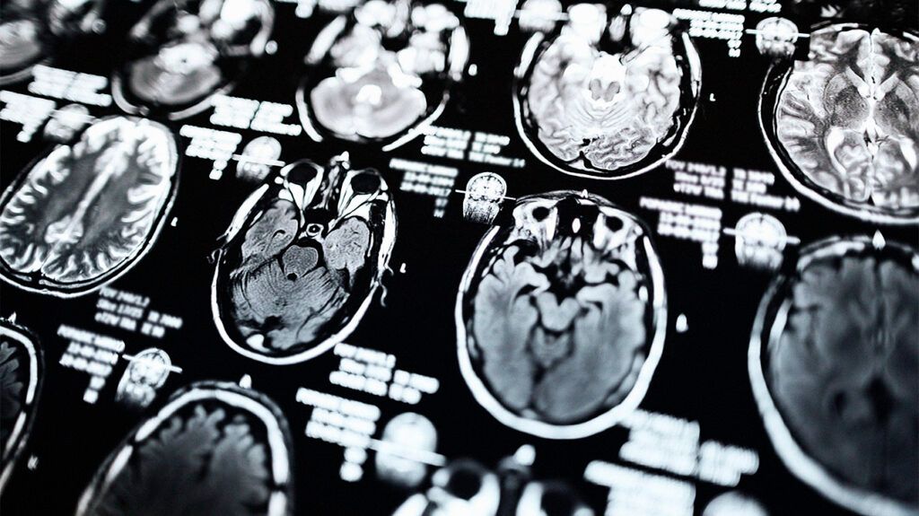
- Scientists may be able to predict disease progression in people with Parkinson’s disease using MRI scans.
- They say structural and functional connections in the brain may help predict gray matter atrophy and progression of the disease.
- Experts say the results of the study could help improve clinical trials for Parkinson’s disease.
MRI scans may help map the functional and structural connections in the brain and allow scientists to predict patterns of atrophy in people with Parkinson’s disease, according to a study published today in Radiology, a journal of the Radiological Society of North America.
In their study, researchers used MRI data from 86 people with mild Parkinson’s disease and 60 healthy controls to map the brain’s neural connections.
They said this information could help them develop an index of disease exposure and predict brain atrophy.
The researchers reported that having Parkinson’s disease for one and two years correlated with atrophy at two to three years past the baseline. Additionally, the models developed by the researchers were able to predict gray matter atrophy accumulation over three years in certain parts of the brain.
The researchers suggest the findings support the theory that functional and structural connections between brain regions could significantly contribute to the progression of Parkinson’s.
The scientists also indicated that the study results suggest that MRI scans could play a role in preventing or delaying disease progression. The researchers suggest that medical professionals incorporate individual-specific information into the model for optimal effectiveness.
“The most significant takeaway of the study is that the brain’s structural and functional connectome significantly influences gray matter atrophy progression in early Parkinson’s disease,” Massimo Filippi, a senior author of the study and a professor of neurology at the Università Vita-Salute San Raffaele in Italy, told Medical News Today. “The study shows that disease exposure indexes, based on healthy brain connectivity and atrophy severity in [Parkinson’s] patients, can predict future atrophy in specific brain regions. This suggests that connectome mapping could be a valuable tool for understanding and predicting the progression of neurodegenerative changes in [Parkinson’s].”
Although this study’s results may not directly help physicians, the researchers said they can enhance clinical studies on Parkinson’s disease and its treatments.
“Medical professionals can use this information to enhance clinical trials. [Disease exposure]-based models could be instrumental in testing the effects of drugs designed to interfere with the spread of alpha-synuclein,” Filippi said. “By accurately predicting atrophy progression, these models can help identify patients who are most likely to benefit from such treatments, thus reducing the number of participants needed and shortening the duration of trials. This targeted approach can lead to more efficient and effective trials, accelerating the development of new therapies for Parkinson’s disease.”
Additionally, a review published in 2019 explained that while conventional MRI scans are not used to diagnose Parkinson’s, they can differentiate it from secondary causes of parkinsonian symptoms.
Parkinson’s disease is a brain disorder that causes unintended or uncontrollable movements, such as shaking, stiffness, and difficulty with balance and coordination, according to the
The risk of developing Parkinson’s increases with age, with most people developing it after age 60. It is more common in men than in women.
Symptoms include:
- Tremor, mainly when someone is at rest
- Slowness of movement
- Limb stiffness
- Gait and balance issues
- Constipation
- Mood changes, including depression, anxiety, and apathy
- Orthostatic hypotension
- Sleep disorders
- Loss of smell
- Skin issues
- Cognitive impairments
There isn’t a cure for Parkinson’s. However, some medications can help with the symptoms.
Levodopa increases dopamine in the brain. However, it does have side effects, including nausea, vomiting, low blood pressure, and restlessness. The drug carbidopa is often taken alongside levodopa to decrease the side effects.
Other medications that can help increase dopamine include dopamine agonists and enzyme inhibitors.
Amantadine and anticholinergic drugs can help reduce involuntary movements, tremors, and muscle rigidity.
Some people find deep brain stimulation works. For this, electrodes are implanted in the brain and connected to a device implanted in the chest. The device stimulates brain areas to help stop involuntary movements and tremors.
